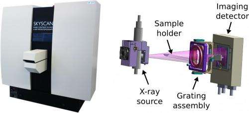
Prototype represents a step toward enhanced soft-tissue tomography

A promising approach for producing medical images with enhanced soft tissue visibility—grating-based x-ray phase contrast—has now advanced from bench-top studies to implementation in an in vivo preclinical computed tomography scanner. A German, Swedish, and Belgian team led by scientists at the Technische Universitaet Muenchen (TUM) published the first experimental results demonstrating the practical potential of this technology, which can significantly improve the contrast in CT scans. This work, reported in the Proceedings of the National Academy of Sciences, could mark a critical step in moving beyond proof-of-concept experiments to applications —including in vivo preclinical imaging with small-animal models in the mid-term future and, in the long term, medical CT scanning.
Conventional x-ray imaging is based on recording the intensity of x-rays behind the imaged object or patient. This method has limitations in medical imaging, especially for examining tissues such as cancer and cartilage, which are hardly visible in conventional images. Novel x-ray technology does not rely solely on the attenuation of x-ray intensity, but also takes advantage of the slightly deflected propagation direction of x-rays. In order to make this effect visible in the so-called phase image, grating-based interferometry is employed, which requires highly precise alignment of x-ray optical components.
Franz Pfeiffer, TUM professor for biomedical physics and head of the research team explains: "For several years we have worked on novel x-ray technology for improved diagnosis in medical imaging. Up until now, we could only investigate excised tissue on bench-top setups in our labs. But now, we took the step of implementing this technology in an in vivo micro-CT scanner. With this step we bring the technology much closer to the hospital, and we hope to make it available for patients in the future."
In a close collaboration with an industrial partner (Bruker microCT / Skyscan), supplied with high-precision x-ray optical gratings from the Karlsruhe Institute of Technology, and with financial support from the European Research Council, the researchers built two prototype scanners. One remains at the industrial partner's site in Belgium, and the other is set up in the Institute of Medical Engineering on TUM's Garching Research Center campus. "The main challenge in implementing the novel x-ray technology in a micro-CT scanner was mechanical stability and resulting disturbances in the x-ray phase images," says TUM doctoral candidate Arne Tapfer, the first author of the PNAS study. "We were able to correct these disturbances with a software algorithm and prove that the correction routine performs accurately." The scientists demonstrated this by quantitatively evaluating the scan of a "phantom" of chemical fluids. Biological tissue was used to assess the potential for biomedical imaging; the novel phase images showed improved visibility of the different tissue components.
"With this new technology we have opened the way for a new generation of preclinical CT scanners," asserts Alexander Sasov, head of the industry partner Bruker microCT—which brought to the project long-standing experience engineering microCT scanners for a wide range of applications.
More information: Experimental results from a preclinical x-ray phase-contrast CT scanner. Arne Tapfer, Martin Bech, Astrid Velroyen, Jan Meiser, Jürgen Mohr, Marco Walter, Joachim Schulz, Bart Pauwels, Peter Bruyndonckx, Xuan Liu, Alexander Sasov, and Franz Pfeiffer. Proceedings of the National Academy of Sciences. PNAS Early Edition for the week of Sept. 10, 2012. www.pnas.org/cgi/doi/10.1073/pnas.1207503109
Journal information: Proceedings of the National Academy of Sciences
Provided by Technical University Munich


