Medical imaging is a vital diagnostic tool, but runs risk of 'false positives'
Medical imaging is a vital tool for the diagnosis of ailments and can help save lives with early detection. But the flip side is a far greater risk of 'false positives', writes David Tan
Following the discovery of X-rays by physicists in 1895, doctors found that this form of electromagnetic radiation could be used to create images of internal structures within our body. Nowadays, there are a plethora of tools on hand to peer at our insides to assess our health, including 3-D imaging such as computed tomography (CT), magnetic resonance imaging (MRI), and ultrasound.
As medical imaging technology keeps improving, and is being used more often for purposes such as cancer screening. Campaigns keep telling us that early detection saves lives, but is this always the case?
We found the PET/MRI enhanced our ability to detect malignant areas
Breast cancer is the most common cancer that affects women in Hong Kong, according to the Hong Kong Breast Cancer Foundation. It cites statistics that show a doubling of female breast cancer diagnoses from 1993 to 2010. On average, eight women are diagnosed with breast cancer every day.
Mammography is a form of X-ray imaging used for screening and detection of masses or calcifications that might indicate breast cancer. The foundation recommends mammography screening every two years for women aged 40 and above, as part of a three-step regimen for early detection of breast cancer.
The survival rate is much higher if cancer is detected early, so early diagnoses can save on medical costs and reduce the impact on the patient.
But the flip side is a risk of over diagnosis. A report from the US in 2010 found that while screening mammograms helped save lives, five to 15 times the number of women were actually unnecessarily diagnosed and treated for cancers that were harmless.
In 2012, a study examining 30 years of data from screening mammography in the US said that while it boosted the detection of early-stage breast cancer, advanced cancer rates were only marginally reduced.
Dr Archie Bleyer and Dr Gilbert Welch said in their report that the study's outcome "suggests there is substantial over-diagnosis, accounting for nearly a third of all newly diagnosed breast cancers, and that screening is having, at best, only a small effect on the rate of death from breast cancer".
X-rays can pass through solid objects, such as our bodies, which is why they are commonly used to image interiors in a non-invasive manner. X-ray imaging works best for dense tissue such as bone, because it absorbs X-rays efficiently.
This means that the level of X-rays reaching the detector is lower than the surroundings, producing a contrast in the final image that makes the tissue visible. Visualisation of internal structures in the body has become an indispensable tool for diagnosis of ailments, from broken bones to tumour masses. CT scanning compiles 2-D X-ray images taken from different angles to build a 3-D image. This has the advantage of producing a more accurate and clearer view of a specific region of interest without superimposing images of other structures.
While X-ray imaging is undoubtedly a useful technology, X-rays are a form of ionising radiation, which can cause damage to DNA in our cells. More recently-developed imaging techniques have moved away from using ionising radiation.
For example, MRI uses a powerful magnetic field to image atoms within the body. Because MRI allows for resolution at the atomic level, it can generate more detailed images than X-rays. MRI is particularly useful for imaging soft tissues in the body, such as the brain, heart and muscles.
Ultrasound uses the reflection of sound waves to produce images, most commonly of fetuses in pregnant women. While ultrasound imaging yields fewer details than CT or MRI imaging, it has the advantage of allowing real-time visualisation of moving structures without using ionising radiation.
Among men, prostate cancer is the third most common cancer in Hong Kong, but this statistic masks the fact that the risk of actually dying from prostate cancer (0.3 per cent) is 10 times lower than the risk of developing this disease (3.2 per cent).
This is because many forms of prostate cancer are non-life threatening. According to Dr Victor Hsue, writing in the September 2013 issue of the "Many cases of prostate cancer do not become clinically evident."
The prostate-specific antigen (PSA) test was introduced about 20 years ago and is the current gold-standard in prostate cancer screening. PSA is a molecule produced by cells in the prostate and PSA levels may rise upon damage to the prostate, indicative of a cancerous growth.
Elevation of PSA levels can be detected five to 10 years before prostate cancer becomes evident, making it suitable for early detection. But PSA levels can also rise in benign conditions, which do not indicate prostate cancer.
Hsue goes on to explain in his report that "Over-diagnosis refers to the detection of tumours by screening that would never be clinically significant." The problems with over-diagnosis include risks associated with the screening process and treatment, as well as potential psychological harm from anxiety after a confirmatory diagnosis.
He writes: "Over-diagnosis is of particular concern because most men with screening-detected prostate cancers have early-stage disease and will be offered aggressive treatment."
Dr Richard Hayward, a London-based neurosurgeon, says the problem lies with the loosening of ties between the medical need for a diagnostic test and the motives for performing the test.
In an opinion article in the , Hayward writes that the advances in medical imaging technology have "allowed such investigations to move tentatively from being purely symptom driven to being non-symptom driven". According to him, performing a scan "just to be on the safe side", can lead to anxiety and fears of a non-existent medical condition.
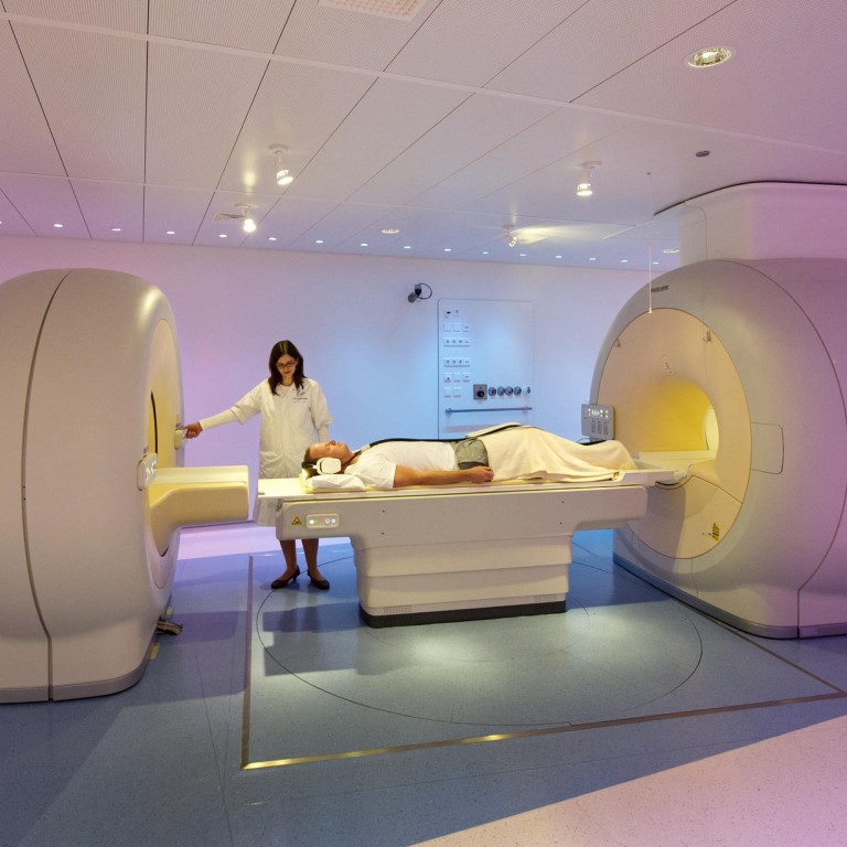
Meanwhile, advances in imaging technology continue to aid more accurate diagnosis. Recently, doctors at the Case Western Reserve University School of Medicine in the US, working in collaboration with Philips Healthcare, successfully combined two forms of imaging technology: MRI with PET (positron emission tomography).
The new PET/MRI hybrid imaging technique marries capabilities of two technologies that are complementary, in order to better visualise both functional and anatomical information and to superimpose this information in a combined digital image.
In so doing, the PET/MRI technology allows doctors to more precisely pinpoint cancer locations and improve the accuracy of disease staging.
The research team says that PET/MRI scanning is useful for staging and treatment planning of colorectal, cervical, uterine, ovarian and pancreatic cancers, as well as in the diagnostic management of paediatric and young adult patients.
Dr Karin Herrmann, a radiologist who led the study, said: "We found the PET/MRI enhanced our ability to detect malignant areas and more accurately and confidently diagnose several types of cancers, potentially providing physicians with the ability to improve treatment planning and better monitoring of the disease."
Dr Peter Faulhaber, another researcher on the team, added: "We are using PET/MRI for pelvic malignancies because there is better soft tissue definition in MRI relative to CT for initial staging."
He believes that the hybrid technique could reduce the number of positive diagnoses made when, in fact, they are negative, a phenomenon known as "false positives".
