

- AI
- Molecular Imaging
- CT
- X-Ray
- Ultrasound
- MRI
- Facility Management
- Mammography
CT Colonography Identifies Pre-Malignant Polyps with Machine-Based Learning Algorithm
Implementing a radiomics-based machine learning algorithm allows CT colonography to differentiate between benign and pre-cancerous polyps with high sensitivity and specificity.
Applying a machine-based learning algorithm to images captured with CT colonography makes it possible for radiologists to differentiate between benign and pre-malignant colorectal polyps.
In industrialized countries across the globe, colorectal cancer ranks among the top three most common causes of cancer-related deaths among both men and women. In a study published Feb. 23 in Radiology, a team of investigators from Germany showed how using radiomics can expand CT colonography beyond simply detecting polyps to pinpointing which ones are likely to progress to being cancerous.
“CT colonography does not enable a definite differentiation between benign and premalignant polyps, crucial for individuals risk stratification and therapy guidance,” said the team led by lead study author Sergio Grosu, M.D., a radiologist from University Hospital at Ludwig Maximilian University in Munich. “In this proof-of-concept study, machine learning-assisted CT colonography analysis allowed for differentiation of benign and premalignant colorectal polyps at high diagnostic performance associated with the histopathologic reference standard.”

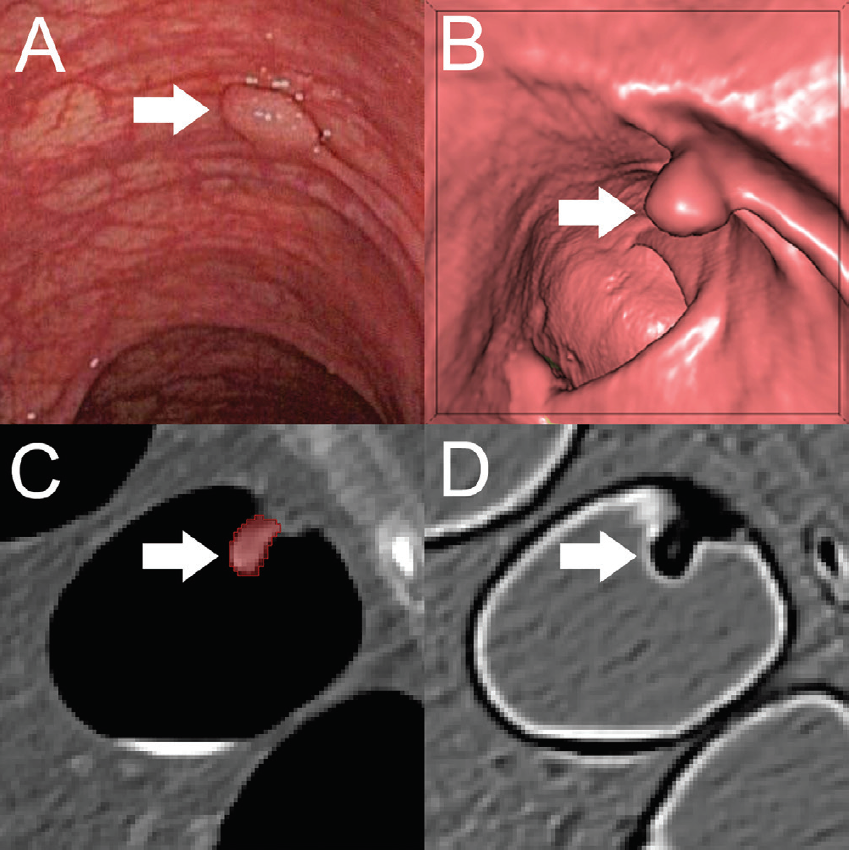
 in the descending colon of a 78-year-old woman. B, Virtual fly-through three-dimensional reconstructions were used for exact polyp localization. C, Manual polyp segmentation was performed in multiplanar two-dimensional CT colonography images. D, CT colonography images were preprocessed for image feature extraction by application of a dedicated filter.
Courtesy: RSNA")
A, Optical colonoscopy and, B–D, CT colonography of a 9-mm polyp (arrow) in the descending colon of a 78-year-old woman. B, Virtual fly-through three-dimensional reconstructions were used for exact polyp localization. C, Manual polyp segmentation was performed in multiplanar two-dimensional CT colonography images. D, CT colonography images were preprocessed for image feature extraction by application of a dedicated filter.
Courtesy: RSNA
For nearly 20 years, CT colonography has gained popularity as a non-invasive screening method for colorectal cancer. It performs equally as well as optical colonoscopy (OC) in visualizing polyps that are at least 6 mm large in asymptomatic patients, and it is also provides better images of complex anatomical cases for which colonoscopy is frequently ineffective.
But, Grosu’s team investigated whether it was possible to push the screening method’s diagnostic value even further to potentially allow for pre-cancerous polyp identification.
To reach this goal, the researchers created a machine learning algorithm that uses radiomics to extract quantitative image features to predict whether the polyps would further develop into cancer. They applied this algorithm to CT colonography images taken in a group of asymptomatic patients who were at average colorectal cancer risk. They, then, trained the algorithm on 169 CT segmentations from 107 colorectal polyps from 63 patients and tested it on 118 segmentations from 77 polyps from 59 patients.

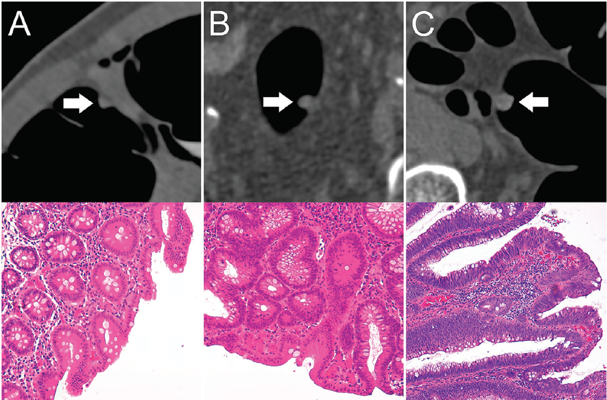
 in the training set. Bottom: Corresponding histopathologic work-up. (Hematoxylin-eosin staining; original magnification 320.) A, An 8-mm hyperplastic polyp in the ascending colon of a 54-year-old woman with hyperplastic epithelia. B, An 8-mm tubular adenoma in the sigmoid colon of a 68-year-old man with tubular growth pattern and elongated nuclei. C, An 11-mm tubulovillous adenoma in the rectum of a 73-year-old man with tubulovillous growth pattern and elongated nuclei.
Courtesy: RSNA")
Top: Axial CT colonography images show representative colorectal polyps (arrow) in the training set. Bottom: Corresponding histopathologic work-up. (Hematoxylin-eosin staining; original magnification 320.) A, An 8-mm hyperplastic polyp in the ascending colon of a 54-year-old woman with hyperplastic epithelia. B, An 8-mm tubular adenoma in the sigmoid colon of a 68-year-old man with tubular growth pattern and elongated nuclei. C, An 11-mm tubulovillous adenoma in the rectum of a 73-year-old man with tubulovillous growth pattern and elongated nuclei.
Courtesy: RSNA
Using random forest analysis, the team determined that their radiomics-based machine learning method allowed for non-invasive differentiation of benign and pre-malignant colorectal polyps with 82-percent sensitivity and 85-percent specificity. The area under the curve (AUC) was excellent, they said, at 0.91.
When the team examined the method’s performance based on the size of the polyps detected, they found that for polyps between 6 mm and 9 mm, the AUC was 0.87 – it was 0.90 for polyps that were at least 10 mm in size. The team also found that the most important image feature for decision making was quantifying first-order gray level histogram statistics.
These results fall in line with existing studies, the team said, but their findings add to the knowledge base around the use of CT colonography for polyp identification because they examined polyps that were smaller than 8 mm. In addition, their algorithm was trained on a balanced training set, and it was validated with an external multi-center test set.
Overall, Grosu said, incorporating a machine learning-derived algorithm can make CT colonography an even better colorectal cancer screening tool.
“Adding machine learning-assisted image analysis to conventional, radiological image reading could further improve the clinical significant of CT colonography-based colorectal cancer screening by allowing for a more precise selection of patients eligible for subsequent polypectomy,” he said. “Furthermore, machine learning-assisted CT colonography image analysis potentially provides the gastroenterologist with a road map of pre-malignant polyps eligible for OC-guided resection, particularly in patients with a high burden of colorectal polyps, such as in familial adenomatous polyposis.”
Still, he said, additional larger studies are needed to further validate the findings and refine the algorithm, making it better suited for the clinical environment.
 Technology Provide a Viable Alternative to X-Rays for Aortic Procedures?")


Study Reveals Benefits of Photon-Counting CT for Assessing Acute Pulmonary Embolism
April 23rd 2024In comparison to energy-integrating detector CT for the workup of suspected acute pulmonary embolism, the use of photon-counting detector CT reduced radiation dosing by 48 percent, according to newly published research.


FDA Approves Fluorescence Imaging System for Detecting Residual Breast Cancer
April 18th 2024The combination of the optical imaging agent Lumisight and the fluorescence imaging device Lumicell Direct Visualization System, collectively known as LumiSystem, reportedly offers 84 percent accuracy with real-time detection of residual breast cancer after lumpectomy procedures.


