Radiology - Technology Information Portal
Monday, 6 May 2024
Info Sheets Out- side     | 'Resolution' p5 Searchterm 'Resolution' found in 5 terms [ • ] and 22 definitions [• ]Result Pages : •
(FWHM) The full width at half maximum is a parameter to characterize the width of a peak on a graph. In nuclear medicine, the FWHM is used to determinate the energy resolution of gamma camera systems. •
(FWTM) The width of a Gaussian or Lorentzian type curve at the tenth of its maximum height (full width at half maximum (FWHM)) and a quality measure of the sharpness of a peak (frequency peak, resolution peak) used less frequently than the FWHM measure.
•  The Koning CT, a Cone Beam CT, provides high spatial and true isotropic resolution at lower dose levels than current CT units. The Koning CT for Breast was introduced at RSNA 2006 and is not cleared for sale in the USA.
Related Product Lines:
CT Systems: 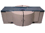
Contact Information
MAIL
Koning Corporation
Lennox Tech Enterprise Center 150 Lucius Gordon Drive, Suite #112 West Henrietta New York 14586 USA
PHONE
+1-585-214-2459
FAX
+1-585-272-0054
ONLINE
CONTACT INFO PAGE
• (MPI) The myocardial perfusion scan is the most common nuclear medicine procedure in cardiac imaging and allows assessing the blood-flow patterns to the heart muscles. The comparison of the radiopharmaceutical distribution after stress and at rest provides information on myocardial viability and cardiac perfusion abnormalities. ECG-gated myocardial perfusion imaging allows the assessment of global and regional myocardial function such as wall motion abnormalities. The diagnostic accuracy of myocardial perfusion scintigraphy (also abbreviated MPS) allows reliable risk stratification and guides the selection of patients for further interventions, such as revascularization. MPI also has particular advantages over alternative techniques in the management of a number of patient subgroups, including women, the elderly, and those with diabetes. The use of this type of cardiac scintigraphy is associated with greater cost effectiveness of treatment, in terms of life-years saved, particularly in these special patient groups. Myocardial perfusion scintigrams are acquired with a gamma camera. Single photon emission computed tomography (SPECT) is preferred over planar imaging because of the three dimensional nature of the images and their superior contrast resolution. Common MPI radiopharmaceuticals, approved by the U.S. Food and Drug Administration (FDA) include: Tl-201 and the Tc-99m-labeled radiopharmaceuticals, such as sestamibi, tetrofosmin, and teboroxime for single-photon imaging. Rb-82 is used for positron emission tomography (PET) imaging. See also Gated Blood Pool Scintigraphy, Myocardial Late Enhancement, Cardiac MRI and Echocardiography. Further Reading: News & More:
•
(NAA) Neutron activation analysis is a very sensitive analytical technique to determine even very low concentration of chemical elements, trace elements for example, in small biological samples. NAA becomes commercial available in the USA in 1960. In the activation process stable nuclides in the sample, which is placed in a neutron beam (neutron flux, 90-95% are thermal neutron with low energy levels under 0.5 eV), will change to radioactive nuclides through neutron capture (artificial radioactivity). These radioactive nuclides decay by emitting alpha-, beta-particles and gamma-rays with a unique half-life. Qualitative and quantitative analysis of the sample is done with a high-resolution gamma-ray spectrometer. NAA is subdivided into the following techniques:
•
•
Prompt Gamma NAA (PGNAA): gamma rays are measured during neutron activation. For detection of elements with a rapid decay.
•
Delayed Gamma NAA (DGNAA): conventional detection after the neutron activation.
•
•
Instrumental NAA (INAA): automated from sample handling to data processing. Analyzes simultaneously more than thirty elements in most samples without chemical processing.
•
Radiochemical NAA (RNAA): After neutron activation the sample is chemically refined for better analysis.
Further Reading: Basics:
News & More: Result Pages : | Share This Page  Look Ups |
Radiology - Technology Information Portal
Member of SoftWays' Medical Imaging Group - MR-TIP • Radiology-TIP • Medical-Ultrasound-Imaging
Copyright © 2008 - 2024 SoftWays. All rights reserved.
Terms of Use | Privacy Policy | Advertising
Member of SoftWays' Medical Imaging Group - MR-TIP • Radiology-TIP • Medical-Ultrasound-Imaging
Copyright © 2008 - 2024 SoftWays. All rights reserved.
Terms of Use | Privacy Policy | Advertising
[last update: 2023-11-06 02:01:00]